ASPIRA-aXA
Pseudophakic reliability for you and your patients
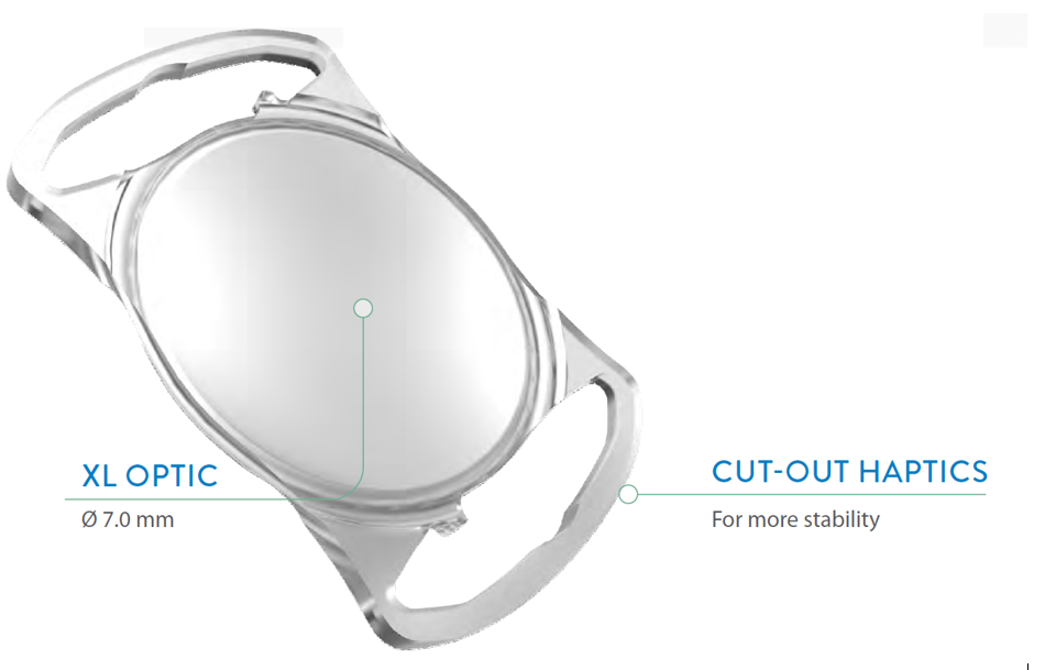
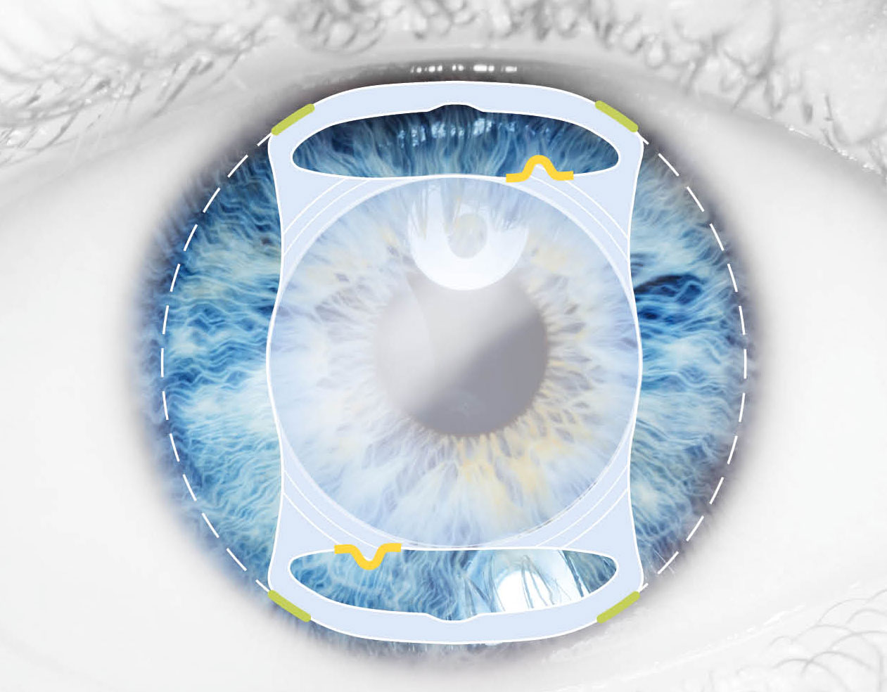
The innovative XL optic of the ASPIRA-aXA combines the advantages of a 7.0 mm optic with the stability of the new cut-out haptic design. This posterior-chamber IOL an be conveniently implanted using small-incision technology while adhering to surgical routine.
Overview of topics
Monofocal ASPIRA-aXA
Personal
Contact
| Type | Monofocal 1-piece posterior chamber lens, foldable |
| Optic diameter | 7.0 mm |
| Total diameter | 11.0 mm |
| Material | Glistening-free, hydrophilic acrylic, UV blocker |
| Optic features | Aspherical anterior surface, aberration-free, 360° LEC barrier |
| Haptic design | Cut-out haptics |
| Constants for IOL calculation | Please click here |
| XL diopter range ASPIRA-aXA | Preloaded SAFELOADER® 10.0 to 30.0 D in 0.5 D steps Compact Line |
| eIFU | Please click here: ASPIRA-aXA Please click here: SAFELOADER® |
| Injector system | Please click here |
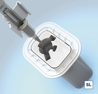
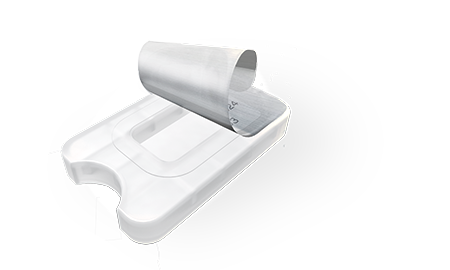
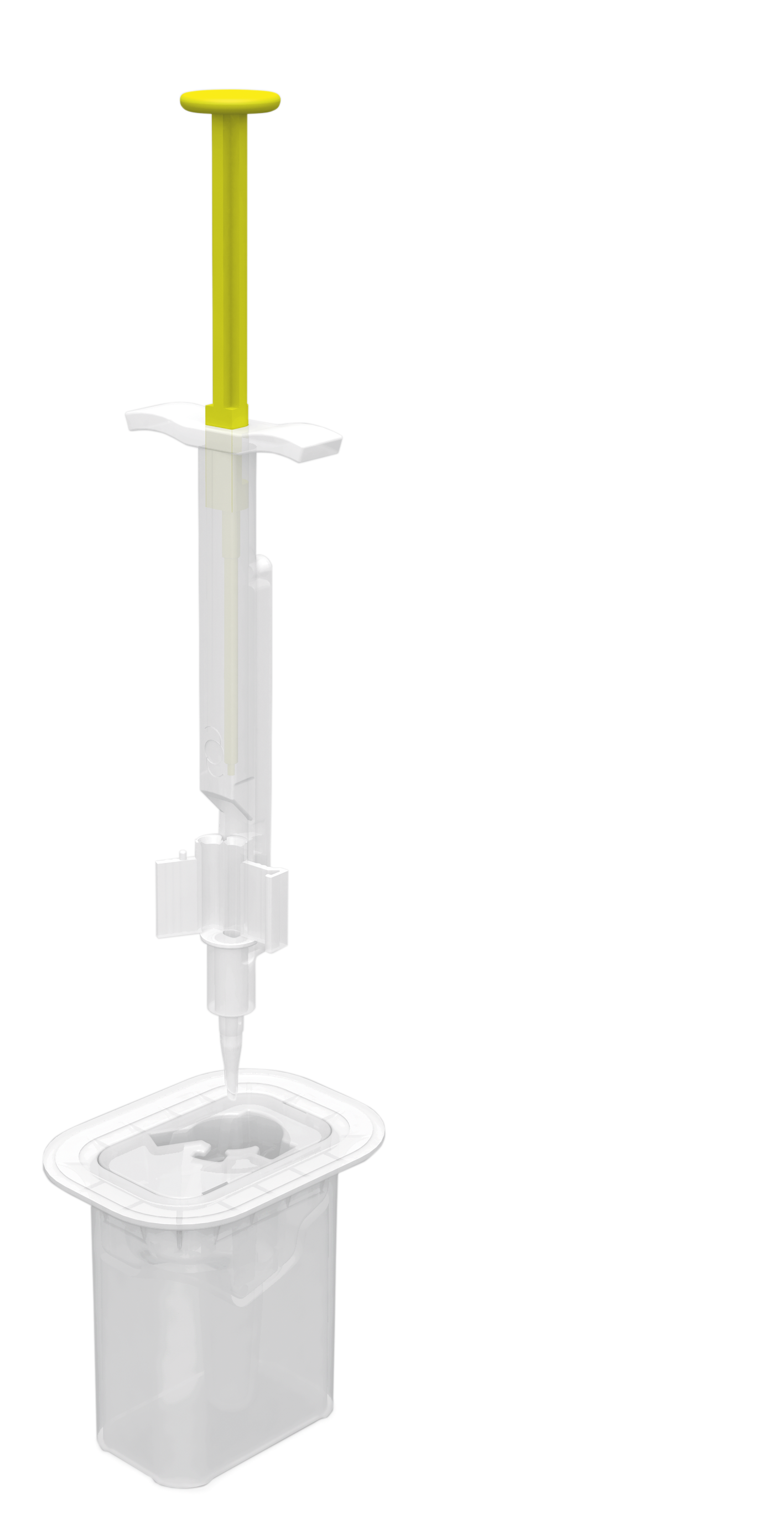
Preloaded implantation system
SIMPLE. INTUITIVE. FAST.
SAFELOADER®
ASPIRA-aXA
THE EXTENDED 7.0 mm XL-OPTIC
For standard cataract surgery and especially for patients with large pupils, traumatic mydriasis or iris defects
For patients at increased risk of retinal diseases or need for combined vitreoretinal surgery
EASY TO INTEGRATE INTO THE ROUTINE
FOR AN UNTROUBLED VISUAL OUTCOME
PREVENT PHOTIC PHENOMENA
Pseudophakic dysphotopsia is the most important dissatisfier for patients after successful cataract surgery.
BENEFIT FROM A SMOOTH ADAPTION PHASE
PREVENT PERSISTENT DYSPHOTOPSIA
Bag-to-bag IOL exchange with the 7.0 mm Aspira-aXA showed complete resolution of dysphotopsia in almost all patients. An IOL exchange with a wide optic diameter IOL seems a promising surgical treatment for dysphotopsia.
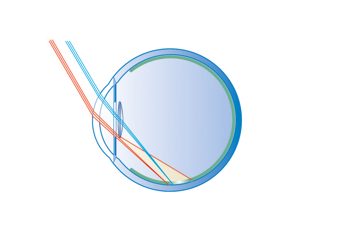
Simulated beam guidance with a 6.0 mm optic
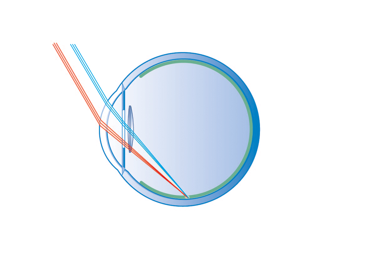
Simulated beam guidance with a 7.0 mm optic
The Aspira-aXA with the 7.0mm XL-optic reduces positive and negative dysphotopsia effectively.
A model
FOR THE DEVELOPMENT OF NEGATIVE DYSPHOTOPSIA
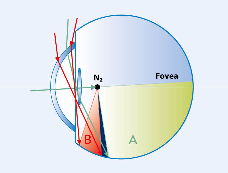
Numerous ray tracing analyses using standard IOLs show a non-illuminated region of the peripheral nasal retina. This “shadow” is caused by the reduced optic diameter and the central thickness of the artificial IOL compared to the natural lens.
Schematic drawing modified according to Holladay
2.5 mm pupil, 6.0 mm IOL
The retinal image is generated by light rays refracted by the IOL optic (areal A), as well as by light rays that hit the retinal periphery (area B) either directly or indirectly refracted by the IOL edge. The unilluminated gap between these two areas can lead to the perception of negative dysphotopsia.
THE PANORAMIC IOL
THINKING ONE STEP AHEAD
THE SOLUTION FOR VITREORETINAL SURGERY
AN INVESTMENT IN THE FUTURE
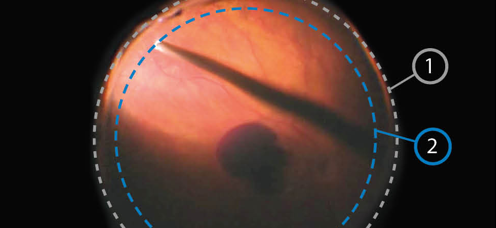
EXCELLENT INTRAOPERATIVE VIEW
Intraoperative fundus image* (1) Edge of the XL optic (2) Theoretical optic edge of a 6.0 mm IOL
* Courtesy of Univ.-Prof. Dr. M. Bolz, Linz
Dr. J. Schrecker, Glauchau, Germany
“We have been using Aspira-aXA for several years – especially in combination with vitreous surgery.
The particular advantage for me is the excellent stability of the IOL position and the vision, especially when using gas and oil tamponades. Even after almost two hundred implantations, no abnormalities, such as calcification or deposits of the hydrophilic material, occurred.“
Personal Statement, 2023
THE PANORAMIC IOL
EXPERTS RECOMMEND
Prof. W. Sekundo, Marburg
“The large ASPIRA-aXA optic has proven itself in phaco-vitrectomies because it allows an edge-free view into the outer retinal periphery.”
Personal Statement, 2018
Univ.-Prof. Dr. Matthias Bolz, MD, Linz
„The view into the periphery is not only important for standard funduscopy, but also for diagnostics, fluorescence angiography and above all for retinal surgery.”
Die ASPIRA-aXA aus Sicht eines Retinologen. Presentation DOC (Internationaler Kongress der Deutschen Ophthalmochirurgen), HumanOptics Booth Lecture, Nuremberg, 2018.
Y. Takamura, MD, PhD.
“The creation of a larger capsulorhexis with implantation of a 7.0 mm optic IOL contributed to a larger anterior capsule opening after cataract surgery in patients with DM [diabetes mellitus].”
Takamura, Y., et al. Large capsulorhexis with implantation of a 7,0 mm optic lens during cataract surgery in patients with diabetes mellitus. J Catract Refract Surg 2014; 40(11):1850-1856.
SAFE POSITIONING
STABLE REFRACTION
Dr. E. Becker, Oranienburg, Germany
“Aspira-aXA combines tilt-free fit, excellent A-constant fidelity with optical precision,
and its size makes it the number one monofocal IOL for eyes with a WTW greater than 12.0 mm and/or large pupils.”
Personal statement, 2023
CLINICALLY CONVINCING
Effective Reduction of Dysphotopsia
Excellent Stability
Highest Level of Patient Satisfaction
| Refraction | Postoperative CDVA stable over time: After 1 month, median 0.00 logMAR; -0.10 bis 0.22 After 1.5 years, median 0.00 logMAR; -0.10 bis 0.10 |
| Mean Tilt | <5.5°, stable in the follow-up period |
| Mean Decentration | <0.2 mm, stable in the follow-up period |
| Median Rotation | 1.8° one week postoperatively No significant changes between surgery and 1.5-year follow-up |
Choose
– AS DESIRED.
7.0 mm optic – preloaded
SAFELOADER®
SIMPLE. INTUITIVE. FAST.
The contactless preloaded implantation system offers a maximum of safety combined with its intuitive, easy handling.
Click here to see the application video
Reports from clinical application
The space-saving model
COMPACT LINE
For space-saving storage even when space is at a premium – ideal for use in the inpatient sector
Premium IOL ASPIRA-aXA
Your advantages of the premium IOL platform
Clinical performance
What do the experts say?
Dr. E. Becker/ M. Bonsemeyer, Oranienburg
“The ASPIRA-aXA with its enlarged XL optic represents a promising approach to minimize dysphotopsia.”
Piskula, M. Becker, E. (2018). Reduzierte pseudophake Dysphotopsien durch vergrößerte IOL-Optik. Presentation DGII 2018.
Prof. G. Duncker, Halle
“The patient response has been overwhelmingly positive.”
Personal Statement, 2018
Application
Publications
Reports from clinical application
Pilger D., Bertelmann E., Brockmann T, et al.
Postoperative Lens Rotation of a 7.0 mm Optic IOL with Plate Haptics
Int J Ophthal Vision Res. 2021 Dec 07;5(1): 014-020.
Language: English
Borkenstein AF, Borkenstein EM
Efficacy of Large Optic Intraocular Lenses in Myopic Eyes with Posterior Segment Pathology
Ophthalmol Ther. 2021 Nov 27.doi: 10.1007/s40123-021-00433-3. Epub 2021 Nov 27.
Language: English
Schrecker J, Seitz B, Langenbucher A
[Performance of a new 7 mm intraocular lens with follow-up over 1.5 years]
Ophthalmologe. 2021 Oct 5.doi: 10.1007/s00347-021-01504-3. Online ahead of print.
Language: German
Bonsemeyer MK, Becker E, Liekfeld A
Dysphotopsiae and functional quality of vision after implantation of an intraocular lens with a 7.0 mm optic and plate haptic design
J Cataract Refract Surg. 2021 Jun 28.doi: 10.1097/j.jcrs.0000000000000735. Online ahead of print.
Language: English
Wendelstein J, Laubichler P, Fischinger I, et al.
Rotational Stability, Tilt and Decentration of a New IOl with a 7.0 mm Optic
Current Eye Research 2021 Jun;14
Click here for full report
Language: English
Borkenstein A. F., Borkenstein E. M.
Creating Hybrid Monovision with 7.0 mm XL Optic and High-Add AMD Intraocular Lenses (XL-MAGS) in a Patient with Retinitis Pigmentosa
Case Rep Ophthalmol 2019;10:304-311; doi: 10.1159/000503093
Click here for full report
Language: English
ASPIRA-aXA
Media reports
This post is also available in: German
OUR
CUSTOMER SERVICE
YOUR
QUESTIONS
Our experts support you with your questions at any time.


